
Ultrasonography
| This factsheet is for people who are having an ultrasound scan, or who would like information about it. | |
| An ultrasound is a procedure that uses sound waves to produce an image of the inside of your body (or part of your body). | |
| You will meet the sonographer or radiologist carrying out your procedure to discuss your care. It may differ from what is described here as it will be designed to meet your individual needs. | |
 |
About ultrasound |
|
Preparing for an ultrasound |
|
What happens during an ultrasound |
|
What are the risks? |
| About ultrasound | |
| You may need to have an abdominal ultrasound to help your doctor identify problems with your liver, gallbladder, pancreas, spleen or kidneys. Pelvic ultrasound can help your doctor identify problems in your lower abdominal and pelvic organs, such as your bladder. | |
| In women, a pelvic ultrasound may be used for a number of reasons, including those listed below. | |
|
To find the cause of pelvic pain, heavy or painful periods or other abnormal vaginal bleeding. |
|
To look for any unusual growths in the ovaries or womb, such as ovarian cysts, fibroids or cancer. |
|
To find the cause of infertility, such as damaged fallopian tubes or endometriosis. |
|
To monitor the effects of fertility treatment. |
|
To check fetal development during pregnancy. |
| Sometimes, ultrasound is used to help guide procedures, such as taking a biopsy (a small sample of tissue). It can also help check for blood clots or narrowing of blood vessels. This is done using a Doppler ultrasound, which monitors flow in blood vessels. The procedure is similar to having a standard ultrasound (see our common questions for more information). | |
| Ultrasound is usually performed by a sonographer. Sonographers are technicians specially trained in taking ultrasound scans. Ultrasound scans can also be performed by a radiologist (a doctor who specialises in using imaging methods to diagnose medical conditions). | |
Ultrasound scans are usually done as out-patient procedures in hospital. Please read your appointment letter for instructions on how to prepare for your scan. The instructions will vary depending on your examination.
For some scans you may be asked to fast for a number of hours beforehand, whereas for others you may need to drink water an hour beforehand. A full bladder helps to lift your large bowel out of the pelvis so it’s easier to examine your pelvic organs.
Your sonographer will discuss with you what will happen before, during and after your procedure, and any pain you might have. This is your opportunity to understand what will happen, and you can help yourself by preparing questions to ask about the risks, benefits and any alternatives to the procedure. This will help you to be informed, so you can give your consent for the procedure to go ahead, which you may be asked to do by signing a consent form.
What happens during an ultrasound
The scan usually takes 10 to 15 minutes and you may be asked to wear a gown.
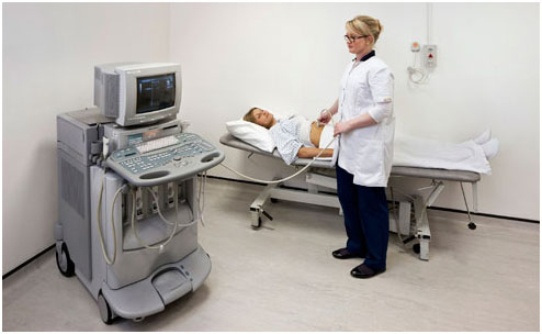
The ultrasound scanner looks a bit like a home computer system. There is a hard-drive, keyboard and a display screen, and a hand-held sensor. The sensor sends out sound waves and picks up the returning echoes. Pictures of the inside of your body are displayed on the screen. These pictures are constantly updated, so the scan can show movement.
Depending on your medical condition you may have a scan of your abdominal or pelvic organs, or both.
Abdominal scan
You will usually need to lie on your back on a couch. Your sonographer will apply gel to the skin on your abdomen (tummy) over the area to be examined. The gel allows the sensor to slide easily over your skin and helps to produce clearer pictures. Your sonographer will hold the sensor firmly against your skin and move it over the surface.
 |
|
||||||||||||||||
| Abdominal ultrasound | |||||||||||||||||
For the transvaginal and transrectal scan, a protective cover is placed over the sensor and it’s well lubricated. The procedure isn’t painful, although it may cause slight discomfort. If you have a latex allergy, tell your sonographer so that he or she uses a suitable cover.
After an ultrasound, your sonographer will wipe the gel from your skin and you will usually be able to go home when you feel ready.
Getting the results
The details of your scan may be explained to you straight after the examination. Alternatively, your results may be sent in a report to the doctor who requested your scan. This can take several days to reach your doctor.
What are the risks?
Ultrasound examination is safe. It doesn’t use radiation and so carries none of the risks associated with this.
Standard diagnostic ultrasound has no side-effects. You may feel slight discomfort as the sensor is pressed against the area being examined, especially if this area is tender.
For answers to frequently asked questions on this topic, see FAQs.
For sources and links to further information, see Resources.